PCR, QPCR, documentazione gel, ELISA / RIA, citometria a flusso, biologia cellulare, purificazione di WB, IHC, DNA / RNA
Down Syndrome Detection
General information
What is Down syndrome?
The most famous and frequent chromosome disorder is mongolism, which is often referred to as Down syndrome because Doctor Langdon Down was the first to describe this syndrome in the medical literature. A syndrome is a disease in which there are abnormalities of different organs. In genetics, and in medicine in general, diseases are often named after the doctor who first described the disease. Down syndrome is one of the most common causes of mental retardation with an average frequency of 1 in 700 births. However, the frequency of Down syndrome increases with the age of the mother. In Down syndrome, the intellectual disability is the main characteristic. This delay is always serious so that patients with Down syndrome learn to walk as a child and cannot go to a regular school, and cannot function independently as an adult. Most patients with Down syndrome also develop dementia from the age of 50 years. About half of Down patients have a congenital heart defect, which often requires complicated surgical procedures. Furthermore, the appearance is also typical: the special eye position that recalls the Mongol breed, has led to the naming Mongolian and Mongolism.
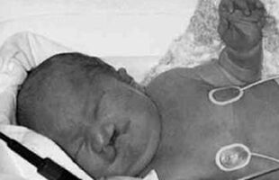
Baby with Down syndrome.
Child with Down syndrome.
Adult with Down syndrome.
Down ptients
What causes Down syndrome?
Down syndrome is caused by an additional third chromosome 21. While everyone has two chromosomes 21, these patients have 3 chromosomes 21 present. It is therefore called a trisomy 21. The cause of a trisomy usually lies in the formation of the germ cells (the egg of the mother or sperm of the father). Normally, the germ cells that come together at fertilization each contain 23 chromosomes with 1 chromosome 21. However, if one of the germ cells has a chromosome 21 too much, the fertilized egg from which the baby is born will have 47 chromosomes, with 3 chromosomes 21.
The risk of a child with Down syndrome increases with the age of the mother.
An average parent couple has a risk of about 1: 200 to a child with a chromosome abnormality. In about half to one third it is a trisomy 21. With the age of the mother, especially from the age of 36, the risk that the egg contains 2 instead of 1 chromosome 21 increases, and so the risk of a child with Down’s syndrome also increases. Therefore, pregnant women who are slightly older (usually from the age of 36) often have an examination during pregnancy (amniocentesis) to rule out that the fruit has a trisomy 21. However, the majority of Down babies are born to young mothers because they have more children than older women. That is why the last 10 years have started screening women of all ages for Down syndrome.
A girl (two X chromosomes) with Down syndrome: there are 47 instead of the normal 46 chromosomes with 3 instead of the normal 2 copies of chromosome 21. It is also called a trisomy 21.
47 Chromosomes
What is heredity?
DNA
Our hereditary material consists of a chemical called nucleic acid. The English translation of nucleic acid is Deoxy Nucleic Acid, which is why it is usually called DNA. The long strand of DNA actually consists of 46 different strands called chromosomes.
All our cells contain chromosomes, which are long strands of hereditary material (DNA). There are only 4 different building blocks of DNA (called nucleic acids or bases): A, C, T & G. In each cell we have about 3 billion bases. These determine the hereditary code because they code for all possible functions of our body.
DNA
Chromosomes
Our cells contain a nucleus that contains the different chromosomes. These chromosomes are long strands of hereditary material (DNA).
A chromosome seen through the microscope: the long strand of DNA is wound up in the chromosome. The offshoots of the coiled DNA strands can be seen at the edge of the chromosome.
Chromosomes
We have 2 copies of each chromosome in each body cell, one of which is from father and one from mother. There are 23 chromosome pairs in each body cell, so a total of 46 chromosomes. However, in the germ cells (egg in the female and sperm in the male) there are only 23 chromosomes, and only one copy of each pair of chromosomes is present. When the 23 chromosomes of the egg combine with the 23 chromosomes of the sperm, a fertilized egg or zygote with 46 chromosomes develops from which the child will develop.
The 46 chromosomes are paired by size. The set of arranged chromosomes is called a karyotype. Each chromosome pair is identified by a number. We have two copies of each chromosome. There are also 2 different sex chromosomes, the X and Y chromosomes (bottom right). This figure shows the karyotype of a man.
Female karyotype: The two sex chromosomes at the bottom right are X chromosomes, and there is no Y chromosome.
Chromosome aberrations
Every normal person has 46 chromosomes in every cell of his body. Individuals with too many or too few chromosomes have a chromosome abnormality. Since there are many different chromosomes, there are also many different chromosome abnormalities. Trisomies are chromosome abnormalities where there is one chromosome too much. These arise when not 23 but 24 chromosomes end up in the egg or sperm cell, so that 1 specific chromosome is present in triplicate instead of in duplicate in the baby. When it comes to chromosome 21, a trisomy 21 therefore arises, with Down’s syndrome as a result. In most cases, these trisomias lead to serious physical and mental abnormalities. The most famous and frequent chromosomal aberration is Down syndrome, also called trisomy 21. Other numeric chromosome abnormalities include trisomy 13 (also called Patau’s syndrome), trisomy 18 (also called Edwards syndrome), and Turner’s syndrome. Trisomy 13 is a very serious chromosomal abnormality in which the children die early. The babies with trisomy 13 also often have cleft lip and palate, brain and heart defects.
Trisomy 13 baby with a cleft lip and palate.
Trisomy 13: There are 3 chromosomes 13, and the total number of chromosomes is 47.
Trisomy 18 is also a serious chromosomal abnormality in which the babies usually die in the first year of life. Characteristics are brain abnormalities, heart defects, a small mouth and abnormal hands with clenched fists.
Trisomy 18 baby with small mouth, split lip, low implanted ears, clenched fist and breathing difficulties.
Trisomy 18 with 47 chromosomes and 3 chromosomes 18.
Which tests are available to screen Down syndrome? First trimester screening
In some Western countries, Downscreening has been performed in the first trimester of pregnancy for several years. This screening test combines fetal ultrasound parameters, in particular crown-trunk length or CRL and nuchal fold measurement or NT, with 2 biochemical parameters: PAPP-A (Pregnancy Associated Placental Protein-A) and ß-HCG (Human Chorionic Gonadotrophin). The ultrasound is best done between 11-13 weeks. The blood sample can be taken at the same time, but it is better to take the blood sample between 9-10 weeks because the results of the test are then known at the time of the NT measurement. This test is not a diagnostic test, but a screening test that only indicates an increased risk for Down’s syndrome. In the case of an abnormal 1st trimester screening test, an amniocentesis or chorionic villus sampling is recommended to rule out a chromosome abnormality in the fetus. Given the time of the screening, a chorionic villus test is still possible at this time of pregnancy, so that the result of the chromosome culture is also known earlier.
Second trimester screening
Maternal screening for Down syndrome can also be performed in the second trimester, and consists of determination of 3 proteins in maternal blood, ß-HCG (Human Chorionic Gonadotrophin), AFP ( a 1 -FoetoProtein) and Free Estriol. Since it concerns a determination of three different components, this test is also called triple test. The triple test is performed in the second trimester of pregnancy between the 14th and 18th weeks. The triple test is not a true diagnostic test, which diagnoses an abnormality in the fetus, but a screening test that only indicates an increased risk for Down’s syndrome. In the event of an abnormal 2nd trimester screening test, amniocentesis is recommended to rule out a chromosome abnormality in the fetus. Given the time of the screening (14th and 18th week), a chorionic villus sampling is no longer appropriate at this time of pregnancy.
How is a prenatal test performed?
Amniocentesis – amniocentesis
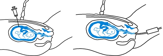
In an amniocentesis, a little (about 20 ml) of amniotic fluid is absorbed from the fetus after a puncture through the abdominal wall and the amniotic membranes. This injection is performed and causes little discomfort or pain to the mother and fetus (comparable to an injection for blood sampling or injection of medicines). An amniocentesis only carries a small risk for pregnancy. In about 1 in 200 cases (0.5%), miscarriage occurs after the injection due to bleeding, infection or amniotic fluid leak. The risk of damage to the fruit or the birth of a child that shows injuries from the amniocentesis is extremely small. Of course, not every miscarriage after an amniocentesis results from this puncture, An amniocentesis is done to obtain both amniotic fluid and fetal cells floating in the amniotic fluid. These amniotic fluid cells are cells of the child, originating from the skin and mucous membranes. Both the amniotic fluid cells and the amniotic fluid can be used for prenatal examination. This research may consist of research on the chromosomes, the metabolism or DNA and on the other hand determination of the alpha foeto protein content (AFP). With each amniocentesis a standard determination of the AFP (to rule out a neural tube defect), and a chromosome examination (to rule out for example that the fetus has Down syndrome) are performed. Sometimes, in addition to the regular chromosome test, a special fast-screening chromosome test is done to rule out some frequent chromosome disorders such as Down syndrome. In many cases, DNA research on cystic fibrosis is now also done. Additionally, research can be performed into specific syndromes at which there is an increased risk. An amniocentesis is preferably performed between the 14th and the 16th week of pregnancy. The results of the AFP test and the fast chromosome test are already known after 3 days. The additional research (full chromosome research, DNA research and biochemical research) usually takes longer (2-4 weeks) because amniotic fluid cells often have to be cultured in the laboratory, which takes time. When an abnormality is found through amniotic fluid examination, the term of pregnancy has often already progressed to the 18th week. If the parents decide to interrupt the pregnancy, this is only possible by inducing the birth with medication.
Amniocentesis: With a needle it is pierced through the abdomen into the amniotic fluid, and about 10-20ml of amniotic fluid (half a glass) is drawn up. Both the amniotic fluid cells floating in the amniotic fluid and the amniotic fluid itself are examined in the laboratory.
The aspirated amniotic fluid is transferred into a tube after the amniocentesis and then examined in the laboratory.
Flake test – chorionic biopsy
In a chorionic villus test, flakes from the fetus are examined (hence the name chorionic villus test). By inserting a suction tube through the cervix, or through a puncture through the abdominal wall and the chorionic membranes, one can aspirate flakes (hence the name chorion bioption). The flakes are offshoots of the placenta, which surrounds the fetus. The puncture of the chorionic villus sampling causes little discomfort or pain to the mother, just like with an amniocentesis. However, the risk of miscarriage is slightly higher than with amniocentesis, and is between 1 and 2% in experienced hands. It is not surprising that this risk is higher than with amniocentesis: The chorionic villus sampling can be used for prenatal testing for the same abnormalities as can be detected with an amniocentesis, with the exception of the AFP content: AFP can only be determined in amniotic fluid and serum. With a chorionic villus test, chromosome, biochemical and DNA research can therefore be performed. Another drawback of the chorionic villus sampling compared to the amniocentesis is the higher risk of miscarriage. But the chorionic villus sampling also has advantages over the amniocentesis. When a DNA test has to be done, it is better to perform a chorionic villus test because flakes are more suitable for DNA research than amniotic fluid. However, the major advantage of the chorionic villus test is that it can be performed earlier than the amniocentesis (11th versus 16th week), while the examinations can often also be carried out directly on the flakes without having to be cultivated. The result of the chorionic villus sampling (12th week) is usually known more than a month earlier than the result of an amniocentesis (18th week). If an abnormality is found during a chorionic villus sampling and the parents wish to have a pregnancy interruption, this can still be done by means of a curettage. This is usually less stressful than inducing labor after an amniocentesis. while the studies can often also be carried out directly on the flakes without having to be cultivated. The result of the chorionic villus sampling (12th week) is usually known more than a month earlier than the result of an amniocentesis (18th week). If an abnormality is found during a chorionic villus sampling and the parents wish to have a pregnancy interruption, this can still be done by means of a curettage. This is usually less stressful than inducing labor after an amniocentesis. while the studies can often also be carried out directly on the flakes without having to be cultivated. The result of the chorionic villus sampling (12th week) is usually known more than a month earlier than the result of an amniocentesis (18th week). If an abnormality is found during a chorionic villus sampling and the parents wish to have a pregnancy interruption, this can still be done by means of a curettage. This is usually less stressful than inducing labor after an amniocentesis. The result of the chorionic villus sampling (12th week) is usually known more than a month earlier than the result of an amniocentesis (18th week). If an abnormality is found during a chorionic villus sampling and the parents wish to have a pregnancy interruption, this can still be done by means of a curettage. This is usually less stressful than inducing labor after an amniocentesis. The result of the chorionic villus sampling (12th week) is usually known more than a month earlier than the result of an amniocentesis (18th week). If an abnormality is found during a chorionic villus sampling and the parents wish to have a pregnancy interruption, this can still be done by means of a curettage. This is usually less stressful than inducing labor after an amniocentesis.
Flake test: a needle is used to pierce the abdomen into the placenta (placenta), and a number of flakes are aspirated. These flakes are examined in the laboratory.
Flake Test
The flakes contain cells from the fetus that can be examined in the laboratory.
What does a genetic advice consist of?
The importance of timely recognition of hereditary disorders is increasingly recognized. That is why people are increasingly referred to a specialist in genetics – the geneticist. Its task is to inform people about hereditary diseases when they are concerned about having or will develop a hereditary disease. The geneticist will also inform completely healthy people about the risks of children with hereditary diseases. Sometimes there are concerns about exposure to harmful substances or the use of medicines before or during pregnancy. If there is already a patient in the family with a disease that may be hereditary, the geneticist will perform the necessary examinations to arrive at a certain diagnosis. The geneticist is also often contacted when an increased risk of Down syndrome has been determined during the screening. The costs of the various forms of heredity testing are largely reimbursed by health insurance, provided that a limited amount of patient contribution is paid.