Cusabio Zaire ebolavirus Recombinant
Description
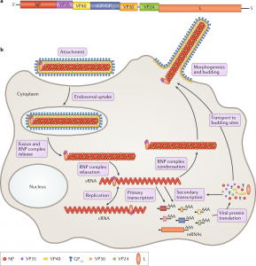
The Zaire ebolavirus Recombinant protein VP35 is an essential component of the viral RNA polymerase complex and also functions to antagonize the cellular type I interferon (IFN) response by blocking the activation of the transcription factor IRF-3. We previously mapped the inhibitory domain of IRF-3 within the C-terminus of VP35. In the present study, we show that mutations that disrupt the IRF-3 inhibitory function of VP35 do not disrupt viral transcription/replication, suggesting that the two functions of VP35 are separable.
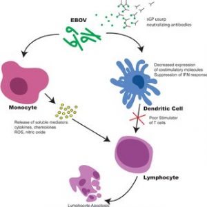
Second, using reverse genetics, we successfully recovered recombinant Ebolaviruses that contained mutations within the IRF-3 inhibitory domain. Importantly, we showed that the recombinant viruses were attenuated for growth in cell culture and that they activated IRF-3 and IRF-3-inducible gene expression at higher levels than that of wild-type VP35-containing Ebola virus. In the context of Ebola virus pathogenesis, VP35 may function to limit the early production of IFN-β and other antiviral signals generated by cells at the primary site of infection, thereby slowing the host’s ability to stop the replication of the virus and induce adaptive immunity.
Purity: greater than 90% as determined by SDS-PAGE.
Target names: NP

Uniprot No.: P18272
Research area: others
Alternative Names: NP Nucleoprotein; PN Ebola; in P; nucleocapsid protein; Protein N
Species: Zaire ebolavirus (Mayinga-76 strain) (ZEBOV) (Zaire Ebola virus)
Source: Yeast
Expression Region: 488-739aa
Mole Weight: 31.1 kDa
Protein length: Partial
Tag Information: N-terminal 6xHis-tagged
Form: Liquid or Lyophilized Powder
Note: We will preferably ship the format we have in stock, however, if you have any special requirements for the format, please remark your requirement when placing the order, we will prepare according to your demand.
Buffer
If the dosage form is liquid, the default storage buffer is Tris/PBS based buffer, 5%-50% glycerol.
Note: If you have any special requirements for glycerol content, please remark it when you order. If the administration form is a lyophilized powder, the buffer before lyophilization is Tris/PBS-based buffer, 6% trehalose, pH 8.0.
Reconstitution
We recommend that this vial be briefly centrifuged before opening to bring the contents to the bottom. Reconstitute protein in sterile deionized water at a concentration of 0.1-1.0 mg/mL. We recommend adding 5-50% glycerol (final concentration) and an aliquot for long-term storage at -20°C/-80°C. Our final default glycerol concentration is 50%. Customers could use it for reference.

Storage Conditions
Store at -20°C/-80°C upon receipt, need to be aliquoted for multiple uses. Avoid repeated cycles of freezing and thawing.
Shelf life
Shelf life is related to many factors, storage condition, buffer ingredients, storage temperature and the stability of the protein itself. Generally, the shelf life of the liquid form is 6 months at -20°C/-80°C. The shelf life of the lyophilized form is 12 months at -20°C/-80°C.
Delivery time: 3-7 business days
Notes: Repeated freezing and thawing is not recommended. Store working aliquots at 4°C for up to one week.
Cells and viruses.
Vero E6 and 293T cells were maintained in Dulbecco’s Modified Eagle’s Medium (DMEM) containing 100 U/ml penicillin, 100 U/ml streptomycins, and 10 mM HEPES buffer (complete) supplemented with fetal bovine serum (FBS) at 10% unless otherwise stated. Huh7 hepatocytes (obtained from Apath, LLC) were cultured in complete DMEM as described above and further supplemented with non-essential amino acids and 10% FBS.
The human monocyte cell line U937 (ATCC CRL-1593.2) was grown in a complete RPMI medium containing 10% FBS. Ebola virus infections were performed in complete DMEM containing 2% FBS. All work with infectious Ebola virus was done inside positive pressure suits in the biosafety level 4 laboratory at the Centers for Disease Control and Prevention. When necessary, samples were quenched for infectivity with 2 × 10⁶ to 5 × 10⁶ rads of cobalt-60 radiation from a Gammacell irradiator.
Western spots.
Cell lysates from the transfections were subjected to sodium dodecyl sulfate-polyacrylamide gel electrophoresis (SDS-PAGE) using the DuPage bis-tris system (Invitrogen). Samples were loaded onto 10% bis-tris gels and electrophoresed for 50 min at 200 V. Proteins were transferred to nitrocellulose membranes using the X-Cell II transfer module (Invitrogen) and then probed with an anti-VP35 monoclonal antibody (1:2000). ) or an antiactin control (1:1,000) (Chemicon). Blots were developed using the Western Breeze Chemiluminescence Detection System (Invitrogen) and exposed to film. Protein expression levels were determined using the AlphaImager EC camera and densitometry software.

